文章信息
- 房煦, 罗军, 高悦, PaulWILLIAMS, 张昊, WilliamDAVISON
- FANG Xu, LUO Jun, GAO Yue, WILLIAMS Paul, ZHANG Hao, William DAVISON
- 梯度扩散薄膜技术(DGT)的理论及其在环境中的应用Ⅱ:土壤与沉积物原位高分辨分析中的方法与应用
- Theory and application of diffusive gradients in thin-films in the environment:High-resolution analysis and its applications in soils and sediments
- 农业环境科学学报, 2017, 36(9): 1693-1702
- Journal of Agro-Environment Science, 2017, 36(9): 1693-1702
- http://dx.doi.org/10.11654/jaes.2017-0454
文章历史
- 收稿日期: 2017-03-28
2. Department of Analytical, Environmental and Geochemistry(AMGC), VUB, Pleinlaan 2, 1050 Brussels, Belgium;
3. Institute for Global Food Security, Queen's University Belfast, Belfast BT9 7BL, United Kingdom;
4. Lancaster Environment Centre, Lancaster University, Lancaster LA1 4YQ, United Kingdom
2. Department of Analytical, Environmental and Geochemistry(AMGC), VUB, Pleinlaan 2, 1050 Brussels, Belgium;
3. Institute for Global Food Security, Queen's University Belfast, Belfast BT9 7BL, United Kingdom;
4. Lancaster Environment Centre, Lancaster University, Lancaster LA1 4YQ, United Kingdom
DGT(Diffusive Gradients in Thin-films)技术自从由Davison和张昊发明以来[1],作为一种金属元素和有机污染物的原位监测和形态分析技术,已经被广泛用于研究实验室和自然条件下待测污染物的环境行为。由于DGT技术基于目标物扩散通量的测量原理,同时其内含的纳米级孔径的扩散层具有对目标物粒径进行选择的能力,当DGT技术应用于土壤及沉积物环境中时,其测量结果不仅体现了间隙水中污染物的浓度以及固相向液相的补给能力[2],同时也排除了土壤和沉积物间隙水中难以被生物直接利用的胶体的贡献[3],从而使得DGT技术作为一种土壤和沉积物中重金属生物有效态浓度的分析工具备受关注。罗军等[4]系统地介绍了DGT的基本原理特性以及在土壤环境中的应用。近年来DGT技术在原位高分辨分析领域获得长足发展,促进了土壤和沉积物重金属生物地球化学的原位研究,为重金属污染控制原理的研究提供了技术支撑。作为DGT未来发展的重要方向之一,DGT技术在土壤和沉积物原位高分辨研究中的应用值得我们进行集中介绍和总结。
土壤和沉积物作为多种化学元素存储和生物化学反应的场所,在元素全球循环中扮演着核心角色,而对元素生物地球化学循环过程的详细解读往往需要了解其在微观尺度下的反应细节。由于土壤和沉积物不具有水和空气的流动性,缺乏有效的混合机制使得空间异质性成为其共有的显著特征。对于土壤,这种异质性主要源自母质矿物、风化程度、生物碎屑的空间分布差异[5]。尤其对于淹水环境下的水稻土,异质性同样受控于竖直方向因氧气消耗和微生物厌氧呼吸带来的氧化还原电位的差异。在沉积物中,氧化还原电位控制的相关元素的纵向分布梯度尤为显著[6-7]。此外,植物根系分泌物、生物扰动和微生物活动会进一步丰富上述环境中的异质性,形成复杂而立体的固液相镶嵌结构。
在已有研究中,微电极[8-11]、逐层采样[12-13]、平面式pH测定[14]以及放射自显影技术[15]已经被用于获取这些环境中溶质空间分布信息。但这些方法大多只能提供一维溶质分布,而且空间分辨率一般也只在厘米至毫米级范围。而DGT技术作为一种研究溶质一维或二维空间分布的工具,其毫米级至亚毫米级的高分辨率特性无疑是理解土壤和沉积物中元素微观地球化学过程的一大进步。同时,作为与DGT技术互补的技术,平板光极通过记录待测物质对发色基团所产生荧光的淬灭,在与DGT技术串联使用时可以进一步实时、空间同步地获取氧气、pH等关键但又难以由后者获取的环境参数的二维分布信息[16-18]。为此,我们将聚焦于DGT技术在土壤和沉积物环境中原位高分辨分析的研究。首先从方法层面概述其对吸附膜和采样装置的要求以及后续分析方法的发展,然后介绍一些重要研究案例,最后对DGT技术在原位高分辨研究中的应用进行总结和展望。
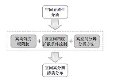
1 基于DGT技术的原位高分辨分析的方法学 1.1 吸附膜的设计为了使用DGT得到土壤或沉积物中目标元素的高分辨率分布信息,一个重要前提就是吸附膜中吸附材料的均匀分布以及其粒径要小于目标分辨率(图 1)。例如当需要进行基于激光剥蚀-电感耦合等离子体质谱仪联用技术(LA-ICP-MS)的亚毫米级分析时,吸附剂颗粒的大小一定要远小于毫米尺度。另外,因为空间异质的溶质向DGT吸附膜扩散过程中会产生程度与扩散层厚度正相关的弥散,进而降低记录到的分辨率[19],所以控制扩散层的厚度对获取有意义的高分辨图像至关重要。
|
| 图 1 DGT原位高分辨分析的思路 Figure 1 High resolution analysis of DGT |
为了提高吸附膜中吸附剂分布的均匀性,有两种思路可供选择:一是在添加吸附剂时使用处理得更细的吸附剂颗粒;二是从原子或分子水平开始将吸附剂组装到吸附胶上。对第一种思路,基于悬浮颗粒试剂亚氨基二乙酸的SPR-IDA吸附膜是典型代表,相比于颗粒大小为200 μm左右的Chelex 100材料,SPR-IDA中0.2 μm左右的细小吸附剂使得由其制成的吸附胶能够胜任微米级的分析[17-18]。
然而更多的用于高分辨分析的吸附胶是通过在胶中由原料直接组装吸附剂而获得的,更具体地说是由原位直接沉淀法制作的基于金属氧化物的吸附膜,包括用于硫离子分析的沉淀型碘化银膜[19-20]和用于含氧阴离子分析的沉淀型氧化铁膜[24-26]与沉淀型氧化锆膜[27]。此类沉淀型吸附膜的制作思路为:首先使用作原料的水凝胶中均匀分布待沉淀的离子,如制作沉淀氧化铁和沉淀氧化锆膜时将制好的水凝胶浸泡在相应的铁盐和锆盐溶液中;然后,将均匀分布好待沉淀离子的水凝胶转移到含有可与之产生沉淀的离子溶液中,使作为吸附剂的沉淀在水凝胶中形成,从而得到吸附剂均匀分布的沉淀型吸附膜[23-24]。
1.1.2 厚度在串联使用DGT和其他单个或多个基于扩散传质的原位技术(如DGT、平板光极等)时,降低总体扩散层厚度从而保证更好的空间分辨率十分关键。除了传统的扩散膜被完全舍弃而以滤膜作为扩散层外,降低吸附膜的厚度也会提升其后诸层的实际分辨能力[28-29]。
1.2 采样装置的设计采样装置设计的一个重要方面就是要保证介质中溶质的二维分布能被真实地记录到DGT中。这不仅包括控制采样过程对介质的扰动,还要能确保所研究的空间范围内溶质的扩散过程得到精确控制。
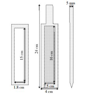
1.2.1 用于沉积物原位分析的装置沉积物的孔隙度或含水率随深度变化的幅度并不陡峭,在DGT探测的空间范围内可认为能够保证一致的扩散条件[25-26]。用于沉积物原位高分辨分析的DGT采样装置设计大都基于标准型DGT探针(图 2)。标准型探针的结构包括一块含有矩形凹槽的塑料支撑板、置于凹槽中的DGT胶层、胶层上覆盖的滤膜以及扣于其最上的一块含有矩形采样窗口的塑料盖板。商品化的标准型DGT探针可通过DGT官网(www.dgtresearch.com或www.dgtresearch.com.cn)购买。该标准型探针既可以用于从原位采集的沉积物柱样[32],也可以通过手动或者水下着陆设备原位插入到沉积物中直接采集目标元素溶质的空间分布信息[24, 33]。虽然这种探针设计最初主要被用于研究目标元素的一维分布,但后续报道表明这种设计同样可以获得元素的二维高分辨分布信息[27, 34-36]。
|
| 自左向右依次为探针盖板、探针主体正面和探针主体侧面 Cap, front view and side view of a DGT probe 图 2 标准型DGT探针结构示意图 Figure 2 Schematic view of a DGT probe |
标准型DGT探针之外,其他尺寸和结构的用户定制或特殊用途的DGT探针同样被广泛采用[37-41]。其中一种由Robertson等[38]开发的大尺寸DGT探针被用于DGT-DET(Diffusive Equilibrium in Thin-films,平衡扩散技术)同时成像,其总尺寸为275 mm×110 mm×14 mm,且有170 mm×80 mm的采样窗口。后续的其他研究中,这种加大型探针也有被再次使用[29, 39-40]。然而由于这种探针底部缺少楔形设计,插入时的阻力以及其对介质的扰动需要额外注意。另一种称为限制型探针的装置中,密集的细条型凹槽设计最初被用于DET空间分辨分析中限制溶质在相邻胶区域间的扩散[39]。由于其能代替高分辨分析中繁琐的切胶步骤,近来同样被用于DGT以分析U、Fe和Mn的毫米级分布[44]。
1.2.2 用于土壤原位分析的装置由于自然土壤含水率一般远低于沉积物,DGT技术用于土壤高分辨原位分析时实验装置往往也有所不同。虽然淹水的稻田土壤依然可以采用用于沉积物的DGT探针,但对于更多的非淹水环境中的土壤,由于其低含水率严重影响溶质的扩散过程,为了在土壤介质中使用DGT技术,需要对采样装置进行改造。其核心就是要保证测量过程中土壤拥有合适而均一的含水率。至今为止,关于用DGT进行土壤原位高分辨的研究大部分是在关注根际土壤环境。
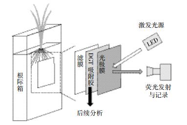
通常待研究的土壤和植物会被置于根际箱中进行培养[25, 28, 45-47](图 3)。通过均匀加水控制土壤含水率到达所需水平,植物培养期间通过控制根际箱的倾斜角度使得植物根系贴壁生长。进行DGT测量时,将覆有滤膜的DGT吸附胶贴于箱板内壁,然后装回根际箱进行测量,既保证了根际土测量所需的含水率,又保证了土壤和根系尽可能贴近DGT膜。
|
| 图 3 DGT用于根际土壤原位高分辨分析的装置图 Figure 3 Design of an experimental device incorporating rhizosphere box, DGT and optode |
DGT技术原位高分辨分析根际土壤和沉积物过程中,为了得到同一目标区域内pH和O2等环境参数的二维分布信息,通常将DGT技术与平板光极技术串联使用[46-47]。实验装置设置如图 3。考虑到DGT吸附胶对平板光极待测物良好的透过性,同时为了避免前者对光极膜荧光信号记录的影响,通常将DGT吸附胶覆盖于平板光极之上,使DGT吸附胶介于待测介质和光极膜之间组成DGT-光极串联结构[47-51]。
1.3 分析方法在选用合适的吸附胶和采样装置完成对待测元素的高精度记录后,选用适当的分析方法读取吸附胶上的二维分布信息将成为DGT高分辨分析中的关键。在针对DET样品的首次原位高分辨分析的尝试中,质子诱导X射线发射光谱(PIXE)被用于获取亚毫米级沉积物中Fe、Mn的分布[52]。但由于PIXE分析昂贵的成本、漫长的时间、仪器的稀缺[53]以及对大多数痕量元素的灵敏度较低[40],基于PIXE的分析方法并不令人满意。
1.3.1 切胶分析法第一个尝试解决上述问题的替代方法是切胶分析法[37]。经过对水凝胶的切割、洗脱后,每小片胶中记录的元素浓度可以通过常用的湿法分析如离子色谱(IC)、石墨炉原子吸收光谱(GF-AAS)、电感耦合等离子体发射光谱(ICP-OES)或电感耦合等离子体质谱(ICP-MS)等准确测定。显然,用于高分辨分析的切胶分析法的关键在于切割得到的胶样品尺寸。
最初的切胶方法为手动把胶切成1 cm长、1~2 mm宽的细小长条[40],但由于过于繁琐且不够精准,便催生出了配备有游标微调器控制的切片机[54],从而能够精确而高效地进行切割。至此,胶样品不仅可以被切成长宽分别为1 cm和1 mm的更小的胶块,提高了竖直方向分辨率,而且还产生了一种切成3 mm×3 mm小胶块式的切割方式,从而更能揭示多元素二维分布的区域化特征和极值点的存在,催生了后续多元素二维成像方面的研究。随后,切胶法所得的分辨率逐渐向亚毫米尺度发展,精确切得的胶块可达450 μm× 450 μm。但由于切胶分析法本身特点决定的对众多细小胶块的后续操作过于耗时,在亚微米级分辨率上的二维高分辨研究还需其他更好的方法。
1.3.2 计算机成像密度法计算机成像密度法(CID)是使用平板扫描仪以图像方式读取DGT胶上溶质的二维分布,并通过计算机图像处理软件分析特定颜色的密度值从而给出待测物浓度的高分辨分析方法。该方法最初被用于测量AgI胶上记录的溶解性硫化物的亚毫米级二维浓度分布[24]。相比于上述的切胶分析法,计算机成像密度法不仅对仪器要求不高,且定量精准(回收率95%,精度5%)[24],极大提高了高分辨二维分析的速度和分辨率,只需一次扫描就能得到理论分辨率238 μm的分布图[24]。
CID分析法显而易见的弊端就是通常只能分析一种元素。这主要由于一般扫描仪只利用三个色彩通道记录颜色,即红色(620~740 nm)、绿色(495~570 nm)和蓝色(450~495 nm),从而使得其对不同分析物各自光谱的解析变得困难。目前使用CID进行二维高分辨分析的方法除了上述的AgI-硫化物系统外,还包括使用菲啰嗪-Fe(Ⅱ)和氧化锆-溶解活性磷(DRP)系统分别对Fe(Ⅱ)和DRP的测定[53, 55-56]。为了解决CID方法分析物单一的问题,一种思路是使用更先进的扫描设备,用更多的色彩通道记录细化的光谱信息,从而使不同分析物的光谱得以分别解析分析,如高光谱成像方法(HSI)[52-53]。然而当同时分析多个元素时,分析就可能变得非常复杂和不准确,这时就需要采用激光剥蚀联用技术。
1.3.3 激光剥蚀联用分析法激光剥蚀-电感耦合等离子体质谱联用技术(LA-ICP-MS)自20世纪80年代出现以来[54-55],已成为当下生物或地质样品成像分析的主流技术[61-63]。由于其基于激光聚焦的微米级剥蚀光斑尺寸和强大的ICP-MS分析系统(图 4),样品的多元素高分辨成像分析技术得到了巨大进步。在2003年首次出现了将LA-ICP-MS应用于DGT样品的高分辨分析的报道,经数据处理后Motelica-Heino等[64]得到了Fe、Mn、Co、Ni和Cu在100 μm分辨率的浓度分布。

|
| 图 4 激光剥蚀电感耦合等离子体质谱分析示意图 Figure 4 Diagram of analysis of DGT using LA-ICP-MS |
将LA-ICP-MS用于DGT吸附胶的高分辨分析,除了LA-ICP-MS固有的多元素同步分析和分辨率的优势,DGT胶的使用还提高了定量分析的准确度。DGT对待测物富集采集的过程可以被看做是一种样品基质纯化和统一化的过程,无论是将DGT放在简单的实验室配置的溶液中还是复杂的环境介质中,最终富集到吸附胶上的待测物都会有一个共同的基质,即凝胶的碳骨架和均匀分布的吸附材料。所以,对于DGT样品分析来说,与样品基质严格匹配的标样通常能够轻易制得,而这是绝大部分基于LA-ICP-MS的固体样品定量分析的难点和关键[60-61]。
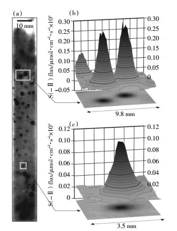
2 应用案例 2.1 沉积物目前使用DGT进行的大部分原位高分辨研究主要是针对沉积物,并取得了一些关键的科学发现。作为一种目前已被广泛认可的地球化学理论,沉积物微生境中金属和硫化物高度区域化的分布和紧密关联的迁移释放就是由DGT的高分辨研究所发现的。因为微生物对硫酸盐等氧化态矿物的还原受控于具有反应活性的有机物,所以硫等许多元素的释放行为都高度依赖于有机质团块的位置、大小以及反应性(图 5)。根据由DGT原位研究获得的这些元素释放的空间规律,人们也重新审视了以前过于简化的一维反应传输模型,开始考虑包含微生境的更加完整的三维反应传输模型[23, 35, 57, 64, 67-70]。
|
| 图 5 (a)扫描得到的AgI胶的灰度图像,其中含53个微生境覆盖了胶6.9%的表面区域(胶尺寸:17.2 mm×141.6 mm),胶表面区域约11%的硫化物的总水平方向净流量可归因于微生境;(b)和(c)圆形微生境的三维图,胶放置在含有取自Esthwaite Water的沉积物的中宇宙中进行。引自Widerlund等[35] Figure 5 (a)Scanned grayscale image of a AgI gel with 53 microniches covering 6.9% of the gel surface area (gel size 17.2 mm×141.6 mm). Approximately 11% of the total horizontal net flux of sulfide on the gel surface area can be attributed to microniches. (b)and(c)Three-dimensional plot of circular microniches. Gel deployed in mesocosm with sediment from Esthwaite Water. Cited from Widerlund et al [35] |
通过使用CID和激光剥蚀高分辨多接收等离子体质谱(LA-HR-MC-ICP-MS)分析DGT样品,准确研究了沉积物间隙水硫化物中硫同位素二维空间(100 μm分辨率)变化特征[71]。发现微生境中δ34S相对背景值高达+20‰,表明如果微生境中这种小尺度的Rayleigh分馏普遍存在(比如对于C、N和Fe),则会对沉积物中元素同位素数据的解读意义重大。
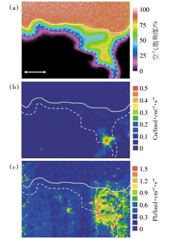
通过串联使用DGT和平面光极技术,Stahl等[51]证明了在沉积物中含有较高氧气浓度的多毛目潜穴壁附近Cu和Pb的局部释放(图 6)。随后Han等[49]进一步结合氧气光极和氧化锆DGT制成复合传感膜,并应用到沉积物中,揭示了其中小尺度异质性的存在。
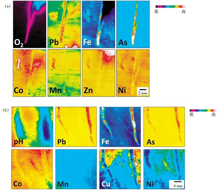
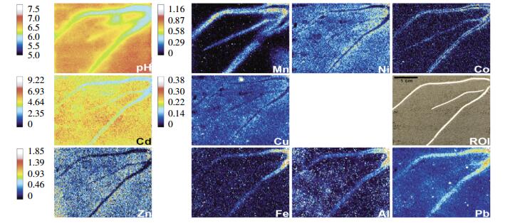
虽然将DGT用于研究土壤和植物根际的研究在2010、2012年才见诸报道[25, 45],但却立即展现了此类应用的广阔前景,并取得了关键的发现。植物根系作为植物与土壤物质交换的通道,其具体过程和状态的阐明对理解植物吸收各种元素有着关键意义,但由于长期缺乏同时测量各种毒性或营养元素有效浓度和关乎元素土壤生物化学过程的含氧量、pH等重要环境参数的原位测定技术,影响植物根系吸收这些元素的过程始终难以精确研究。具有原位高分辨分析能力的DGT技术与平板光极技术的联用克服了之前的困难。通过在根际箱实验中串联使用DGT和平板光极,Williams等[47]首次发现水稻根尖附近存在一个As、Pb和Fe释放的显著提高,同时伴随着氧气富集和pH降低的区域(图 7)。在另一项研究中,Hoefer等[46]同样在柳苗(Salix smithiana)根部附近观测到了更高的Zn和Cd的释放,随后Hoefer等[50]又将pH光极膜和DGT吸附膜整合到同一水凝胶层,从而实现了对柳苗根际土壤pH和金属释放的时空同步成像研究(图 8)。
|
| 图 7 (a)一组水稻根部附近O2、Pb、Fe、As、Co、Mn、Zn和Ni的图像;测量通过纵向放置O2光极-DGT传感器到厌氧土壤中,并围绕一淹没于土壤表面下10 mm的根尖进行;(b)一组生长在同一土壤中水-土界面下10.5 cm深度的水稻根部附近pH和Pb、Fe、As、Co、Mn、Cu、Ni的二维呈现,图像由一pH光极组合一超薄0.05 mm的Chelex DGT获得。引自Williams等[47] Figure 7 (a)Visualization of O2, Pb, Fe, As, Co, Mn, Zn, and Ni around a set of rice roots. Measurements were made by deploying an O2 optode-DGT sensor vertically in the anoxic soil and encompassed a submerged root tip located 10 mm below the soil surface. (b)Two-dimensional representation of pH, Pb, Fe, As, Co, Mn, Cu, and Ni around a different set of rice root grown in the same soil at a depth of 10.5 cm from the soil-water interface. The images were obtained from a pH optode partnered with an ultrathin 0.05 mm Chelex DGT. Cited from Williams et al [47] |
|
| 图 8 Salix smithiana根际的化学成像。每种分析物图像左侧的竖条显示了Cd2+、Zn2+、Mn2+、Cu2+的DGT流量(pg·cm-2·s-1)和pH的标定范围,Fe、Ni2+、Al3+、Co2+、Pb2+则以未经标定、经13C标准化的值显示,并使用同经标定图像相同的颜色体系,即高胶负载值对应亮色(白)而低胶负载值对应暗色(黑)。研究区域(Region of Interest,ROI)为平面光极-DGT放置前拍摄的照片。引自Hoefer等[50] Figure 8 Chemical images of the Salix smithiana rhizosphere. Bars on the left side of each analyte image show the calibration ranges for Cd2+, Zn2+, Mn2+, Cu2+ as DGT flux (pg· cm-2·s-1)and pH. Fe, Ni2+, Al3+, Co2+ and Pb2+ are shown as uncalibrated, 13C-normalized values, utilizing the same color scheme as for the calibrated images with high gel loadings corresponding to bright(white)and low gel loadings corresponding to dark (black)color. The region of interest (ROI)displays a photograph taken prior to PO-DGT deployment. Cited from Hoefer et al [50] |
自DGT技术及其姊妹技术DET诞生以来,对异质性环境介质的原位高分辨研究始终是一个重要的发展方向。为了更准确、更方便地用DGT技术研究介质中待测物的空间分布,DGT的吸附胶、装置等都经过了专门的优化设计;为了更准确方便地读取DGT记录在吸附胶上的溶质分布,多种高分辨测量手段也进行了不断地创新和发展;而为了更科学地解读在土壤和沉积物中DGT测得的结果,一整套的DGT理论被建立和完善起来,同时还开发出了用于模拟计算的计算机软件DIFS模型(详见本系列第一篇[4])。
作为一种被动采样技术,DGT及其相关技术突破性地测量了具有环境意义的元素的有效态浓度,并实现了对土壤和沉积物中重金属的高分辨原位研究。尤其在《水污染防治行动计划》(水十条)、《土壤污染防治行动计划》(土十条)出台及国家对重金属污染和食品安全问题越来越重视的大背景下,DGT技术在沉积物和土壤中对污染物微界面机制的原位高分辨率研究,将为揭示控制重金属污染的关键过程,从而针对性地制定管理控制方案提供重要理论支持。同时,相关的新型高均匀度吸附膜和基于复合材料的重金属-营养盐-环境参数等多指标同步监测技术的开发,以及利用DGT原位高分辨技术对沉积物、根际土壤中关键生物地球化学过程的研究应用,都将作为未来DGT原位高分辨研究的热点而具有广阔的发展空间。
| [1] |
Davison W, Zhang H. In situ speciation measurements of trace components in natural waters using thin-film gels[J]. Nature, 1994, 367(6463): 546-548. DOI:10.1038/367546a0 |
| [2] |
Zhang H, Zhao F J, Sun B, et al. A new method to measure effective soil solution concentration predicts copper availability to plants[J]. Environmental Science & Technology, 2001, 35(12): 2602-2607. |
| [3] |
Van Der Veeken P L R, Pinheiro J P, Van Leeuwen H P. Metal speciation by DGT/DET in colloidal complex systems[J]. Environmental Science & Technology, 2008, 42(23): 8835-8840. |
| [4] |
罗军, 王晓蓉, 张昊, 等. 梯度扩散薄膜技术(DGT)的理论及其在环境中的应用Ⅰ:工作原理、特性与在土壤中的应用[J]. 农业环境科学学报, 2011, 30(2): 205-213. LUO Jun, WANG Xiao-rong, ZHANG Hao, et al. Theory and application of diffusive gradients in thin films in soils[J]. Journal of Agro-Environment Science, 2011, 30(2): 205-213. |
| [5] |
Scheffer F, Schachtschabel P, Blume H, et al. Lehrbuch der Bodenkunde[M]. 2002, 593.
|
| [6] |
Berner R A. Early diagenesis:A theoretical approach[M]. Princeton: Princeton University Press, 1980.
|
| [7] |
Aller R C. Sedimentary diagenesis, depositional environments, and benthic fluxes[J]. Treatise on Geochemistry, 2014, 8: 293-334. |
| [8] |
Stockdale A, Davison W, Zhang H. Micro-scale biogeochemical heterogeneity in sediments:A review of available technology and observed evidence[J]. Earth-Science Reviews, 2009, 92(1): 81-97. |
| [9] |
Kühl M. The benthic boundary layer[M]. New York: Oxford University Press, 2001, 180-210.
|
| [10] |
Weisenseel M H, Dorn A, Jaffe L F. Natural H+ currents traverse growing roots and root hairs of barley(Hordeum vulgare L.)[J]. Plant Physiology, 1979, 64(3): 512-518. DOI:10.1104/pp.64.3.512 |
| [11] |
Taillefert M, Luther G W, Nuzzio D B. The application of electroanalytical tools for in situ measurements in aquatic systems[J]. Electroanalysis, 2000, 12(6): 401-412. DOI:10.1002/(ISSN)1521-4109 |
| [12] |
Wenzel W W, Wieshammer G, Fitz W J, et al. Novel rhizobox design to assess rhizosphere characteristics at high spatial resolution[J]. Plant and Soil, 2001, 237(1): 37-45. DOI:10.1023/A:1013395122730 |
| [13] |
Teasdale P R, Batley G E, Apte S C, et al. Pore water sampling with sediment peepers[J]. Trends in Analytical Chemistry, 1995, 14(6): 250-256. DOI:10.1016/0165-9936(95)91617-2 |
| [14] |
Jaillard B, Ruiz L, Arvieu J C. pH mapping in transparent gel using color indicator videodensitometry[J]. Plant and Soil, 1996, 183(1): 85-95. DOI:10.1007/BF02185568 |
| [15] |
Bhat K K S, Nye P H. Diffusion of phosphate to plant roots in soil[J]. Plant and Soil, 1973, 38(1): 161-175. DOI:10.1007/BF00011224 |
| [16] |
Han C, Ren J, Tang H, et al. Quantitative imaging of radial oxygen loss from Valisneria spiralis roots with a fluorescent planar optode[J]. Science of the Total Environment, 2016, 569/570: 1232-1240. DOI:10.1016/j.scitotenv.2016.06.198 |
| [17] |
Glud R N, Ramsing N B, Gundersen J K, et al. Planar optodes:A new tool for fine scale measurements of two-dimensional O2 distribution in benthic communities[J]. Marine Ecology Progress Series, 1996, 140(1/2/3): 217-226. |
| [18] |
Santner J, Larsen M, Kreuzeder A, et al. Two decades of chemical imaging of solutes in sediments and soils:A review[J]. Analytica Chimica Acta, 2015, 878: 9-42. DOI:10.1016/j.aca.2015.02.006 |
| [19] |
Davison W, Zhang H, Grime G W. Performance characteristics of gel probes used for measuring the chemistry of pore waters[J]. Environmental Science & Technology, 1994, 28(9): 1623-1632. |
| [20] |
Warnken K W, Zhang H, Davison W. Analysis of polyacrylamide gels for trace metals using diffusive gradients in thin films and laser ablation inductively coupled plasma mass spectrometry[J]. Anal Chem, 2004, 76(20): 6077-6084. DOI:10.1021/ac0400358 |
| [21] |
Warnken K W, Zhang H, Davison W. Performance characteristics of suspended particulate reagent-iminodiacetate as a binding agent for diffusive gradients in thin films[J]. Analytica Chimica Acta, 2004, 508(1): 41-51. DOI:10.1016/j.aca.2003.11.051 |
| [22] |
Devries C R, Wang F Y. In situ two-dimensional high-resolution profiling of sulfide in sediment interstitial waters[J]. Environmental Science & Technology, 2003, 37(4): 792-797. |
| [23] |
Stockdale A, Davison W, Zhang H. High-resolution two-dimensional quantitative analysis of phosphorus, vanadium and arsenic, and qualitative analysis of sulfide, in a freshwater sediment[J]. Environmental Chemistry, 2008, 5(2): 143-149. DOI:10.1071/EN07096 |
| [24] |
Teasdale P R, Hayward S, Davison W. In situ, high-resolution measurement of dissolved sulfide using diffusive gradients in thin films with computer-imaging densitometry[J]. Analytical Chemistry, 1999, 71(11): 2186-2191. DOI:10.1021/ac981329u |
| [25] |
Santner J, Prohaska T, Luo J, et al. Ferrihydrite containing gel for chemical imaging of labile phosphate species in sediments and soils using diffusive gradients in thin films[J]. Anal Chem, 2010, 82(18): 7668-7674. DOI:10.1021/ac101450j |
| [26] |
Luo J, Zhang H, Santner J, et al. Performance characteristics of diffusive gradients in thin films equipped with a binding gel layer containing precipitated ferrihydrite for measuring arsenic(Ⅴ), selenium(Ⅵ), vanadium(Ⅴ), and antimony(Ⅴ)[J]. Analytical Chemistry, 2010, 82(21): 8903-8909. DOI:10.1021/ac101676w |
| [27] |
Guan D X, Williams P N, Luo J, et al. Novel precipitated zirconia-based DGT technique for high-resolution imaging of oxyanions in waters and sediments[J]. Environmental Science & Technology, 2015, 49(6): 3653-3661. |
| [28] |
Kreuzeder A, Santner J, Prohaska T, et al. Gel for simultaneous chemical imaging of anionic and cationic solutes using diffusive gradients in thin films[J]. Analytical Chemistry, 2013, 85(24): 12028-12036. DOI:10.1021/ac403050f |
| [29] |
Lehto N J, Davison W, Zhang H. The use of ultra-thin diffusive gradients in thin-films(DGT) devices for the analysis of trace metal dynamics in soils and sediments:A measurement and modelling approach[J]. Environmental Chemistry, 2012, 9(4): 415-423. DOI:10.1071/EN12036 |
| [30] |
Bahr D B, Hutton E W H, Syvitski J P M, et al. Exponential approximations to compacted sediment porosity profiles[J]. Computers and Geosciences, 2001, 27(6): 691-700. DOI:10.1016/S0098-3004(00)00140-0 |
| [31] |
Ehrenberg S N, Nadeau P H. Sandstone vs. carbonate petroleum reservoirs:A global perspective on porosity-depth and porosity-permeability relationships[J]. AAPG Bulletin, 2005, 89(4): 435-445. DOI:10.1306/11230404071 |
| [32] |
Zhang H, Davison W, Gadi R, et al. In situ measurement of dissolved phosphorus in natural waters using DGT[J]. Analytica Chimica Acta, 1998, 370(1): 29-38. DOI:10.1016/S0003-2670(98)00250-5 |
| [33] |
Fones G R, Davison W, Holby O, et al. High-resolution metal gradients measured by in situ DGT/DET deployment in black sea sediments using an autonomous benthic lander[J]. Limnology and Oceanography, 2001, 46(4): 982-988. DOI:10.4319/lo.2001.46.4.0982 |
| [34] |
Pagès A, Teasdale P R, Robertson D, et al. Representative measurement of two-dimensional reactive phosphate distributions and co-distributed iron(Ⅱ) and sulfide in seagrass sediment porewaters[J]. Chemosphere, 2011, 85(8): 1256-1261. DOI:10.1016/j.chemosphere.2011.07.020 |
| [35] |
Widerlund A, Davison W. Size and density distribution of sulfide-producing microniches in lake sediments[J]. Environmental Science & Technology, 2007, 41(23): 8044-8049. |
| [36] |
Bennett W W, Teasdale P R, Welsh D T, et al. Inorganic arsenic and iron(Ⅱ) distributions in sediment porewaters investigated by a combined DGT-colourimetric DET technique[J]. Environmental Chemistry, 2012, 9(1): 31-40. DOI:10.1071/EN11074 |
| [37] |
Krom M D, Davison P, Zhang H, et al. High-resolution pore-water sampling with a gel sampler[J]. Limnology and Oceanography, 1994, 39(8): 1967-1972. DOI:10.4319/lo.1994.39.8.1967 |
| [38] |
Robertson D, Teasdale P R, Welsh D T. A novel gel-based technique for the high resolution, two-dimensional determination of iron(Ⅱ) and sulfide in sediment[J]. Limnology and Oceanography-Methods, 2008, 6(10): 502-512. DOI:10.4319/lom.2008.6.502 |
| [39] |
Fones G R, Davison W, Grime G W. Development of constrained DET for measurements of dissolved iron in surface sediments at sub-mm resolution[J]. Science of the Total Environment, 1998, 221(2/3): 127-137. |
| [40] |
Zhang H, Davison W, Miller S, et al. In situ high resolution measurements of fluxes of Ni, Cu, Fe, and Mn and concentrations of Zn and Cd in porewaters by DGT[J]. Geochimica et Cosmochimica Acta, 1995, 59(20): 4181-4192. DOI:10.1016/0016-7037(95)00293-9 |
| [41] |
Bennett W W, Welsh D T, Serriere A, et al. A colorimetric DET technique for the high-resolution measurement of two-dimensional alkalinity distributions in sediment porewaters[J]. Chemosphere, 2015, 119: 547-552. DOI:10.1016/j.chemosphere.2014.07.042 |
| [42] |
Robertson D, Welsh D T, Teasdale P R. Investigating biogenic heterogeneity in coastal sediments with two-dimensional measurements of iron(Ⅱ) and sulfide[J]. Environmental Chemistry, 2009, 6(1): 60-69. DOI:10.1071/EN08059 |
| [43] |
Pagès A, Grice K, Vacher M, et al. Characterizing microbial communities and processes in a modern stromatolite(Shark Bay) using lipid biomarkers and two-dimensional distributions of porewater solutes[J]. Environmental Microbiology, 2014, 16(8): 2458-2474. DOI:10.1111/1462-2920.12378 |
| [44] |
Docekal B, Gregusova M. Segmented sediment probe for diffusive gradient in thin films technique[J]. Analyst, 2012, 137(2): 502-507. DOI:10.1039/C1AN15701A |
| [45] |
Santner J, Zhang H, Leitner D, et al. High-resolution chemical imaging of labile phosphorus in the rhizosphere of Brassica napus L. cultivars[J]. Environmental and Experimental Botany, 2010, 77: 219-226. |
| [46] |
Hoefer C, Santner J, Puschenreiter M, et al. Localized metal solubilization in the rhizosphere of Salix smithiana upon sulfur application[J]. Environmental Science & Technology, 2015, 49(7): 4522-4529. |
| [47] |
Williams P N, Santner J, Larsen M, et al. Localized flux maxima of arsenic, lead, and iron around root apices in flooded lowland rice[J]. Environmental Science & Technology, 2014, 48(15): 8498-8506. |
| [48] |
Christel W, Zhu K, Hoefer C, et al. Spatiotemporal dynamics of phosphorus release, oxygen consumption and greenhouse gas emissions after localised soil amendment with organic fertilisers[J]. Science of the Total Environment, 2016, 554/555: 119-129. DOI:10.1016/j.scitotenv.2016.02.152 |
| [49] |
Han C, Ren J, Wang Z, et al. A novel hybrid sensor for combined imaging of dissolved oxygen and labile phosphorus flux in sediment and water[J]. Water Research, 2017, 108: 179-188. DOI:10.1016/j.watres.2016.10.075 |
| [50] |
Hoefer C, Santner J, Borisov S M, et al. Integrating chemical imaging of cationic trace metal solutes and pH into a single hydrogel layer[J]. Analytica Chimica Acta, 2017, 950: 88-97. DOI:10.1016/j.aca.2016.11.004 |
| [51] |
Stahl H, Warnken K W, Sochaczewski L, et al. A combined sensor for simultaneous high resolution 2-D imaging of oxygen and trace metals fluxes[J]. Limnology and Oceanography:Methods, 2012, 10(5): 389-401. DOI:10.4319/lom.2012.10.389 |
| [52] |
Davison W, Grime G W, Morgan J A W, et al. Distribution of dissolved iron in sediment pore waters at submillimetre resolution[J]. Nature, 1991, 352(6333): 323-325. DOI:10.1038/352323a0 |
| [53] |
Jézéquel D, Brayner R, Metzger E, et al. Two-dimensional determination of dissolved iron and sulfur species in marine sediment pore-waters by thin-film based imaging, Thau lagoon(France)[J]. Estuarine, Coastal and Shelf Science, 2007, 72(3): 420-431. DOI:10.1016/j.ecss.2006.11.031 |
| [54] |
Shuttleworth S M, Davison W, Hamilton-Taylor J. Two-dimensional and fine structure in the concentrations of iron and manganese in sediment pore-waters[J]. Environmental Science & Technology, 1999, 33(23): 4169-4175. |
| [55] |
Ding S M, Jia F, Xu D, et al. High-resolution, two-dimensional measurement of dissolved reactive phosphorus in sediments using the diffusive gradients in thin films technique in combination with a routine procedure[J]. Environmental Science & Technology, 2011, 45(22): 9680-9686. |
| [56] |
Ding S M, Wang Y, Xu D, et al. Gel-based coloration technique for the submillimeter-scale imaging of labile phosphorus in sediments and soils with diffusive gradients in thin films[J]. Environmental Science & Technology, 2013, 47(14): 7821-7829. |
| [57] |
Cesbron F, Metzger E, Launeau P, et al. Simultaneous 2D imaging of dissolved iron and reactive phosphorus in sediment porewaters by thin-film and hyperspectral methods[J]. Environmental Science & Technology, 2014, 48(5): 2816-2826. |
| [58] |
Manley M. Near-infrared spectroscopy and hyperspectral imaging:Non-destructive analysis of biological materials[J]. Chemical Society Reviews, 2014, 43(24): 8200-8214. DOI:10.1039/C4CS00062E |
| [59] |
Arrowsmith P, Hughes S K. Entrainment and transport of laser ablated plumes for subsequent elemental analysis[J]. Applied Spectroscopy, 1988, 42(7): 1231-1239. DOI:10.1366/0003702884430100 |
| [60] |
Gray A L. Solid sample introduction by laser ablation for inductively coupled plasma source mass spectrometry[J]. Analyst, 1985, 110(5): 551-556. DOI:10.1039/an9851000551 |
| [61] |
Becker J S, Matusch A, Wu B. Bioimaging mass spectrometry of trace elements-recent advance and applications of LA-ICP-MS:A review[J]. Analytica Chimica Acta, 2014, 835: 1-18. DOI:10.1016/j.aca.2014.04.048 |
| [62] |
Koch J, Gunther D. Review of the state-of-the-art of laser ablation inductively coupled plasma mass spectrometry[J]. Applied Spectroscopy, 2011, 65(5): 155A-162A. DOI:10.1366/11-06255 |
| [63] |
Kindness A, Sekaran C N, Feldmann J. Two-dimensional mapping of copper and zinc in liver sections by laser ablation-inductively coupled plasma mass spectrometry[J]. Clin Chem, 2003, 49(11): 1916-1923. DOI:10.1373/clinchem.2003.022046 |
| [64] |
Motelica-Heino M, Naylor C, Zhang H, et al. Simultaneous release of metals and sulfide in lacustrine sediment[J]. Environmental Science & Technology, 2003, 37(19): 4374-4381. |
| [65] |
Pisonero J, Fliegel D, Gu D, et al. High efficiency aerosol dispersion cell for laser ablation-ICP-MS[J]. Journal of Analytical Atomic Spectrometry, 2006, 21(9): 922-931. DOI:10.1039/B603867K |
| [66] |
Kivel N, Potthast H-D, Günther-Leopold I, et al. Modeling of the plasma extraction efficiency of an inductively coupled plasma-mass spectrometer interface using the direct simulation Monte Carlo method[J]. Spectrochimica Acta Part B:Atomic Spectroscopy, 2014, 93: 34-40. DOI:10.1016/j.sab.2013.12.010 |
| [67] |
Pages A, Welsh D T, Teasdale P R, et al. Diel fluctuations in solute distributions and biogeochemical cycling in a hypersaline microbial mat from Shark Bay, WA[J]. Marine Chemistry, 2014, 167: 102-112. DOI:10.1016/j.marchem.2014.05.003 |
| [68] |
Fones G R, Davison W, Hamilton-Taylor J. The fine-scale remobilization of metals in the surface sediment of the North-east Atlantic[J]. Continental Shelf Research, 2004, 24(13/14): 1485-1504. |
| [69] |
Tankere-Muller S, Zhang H, Davison W, et al. Fine scale remobilisation of Fe, Mn, Co, Ni, Cu and Cd in contaminated marine sediment[J]. Marine Chemistry, 2007, 106(1): 192-207. |
| [70] |
Sochaczewski L, Stockdale A, Davison W, et al. A three-dimensional reactive transport model for sediments, incorporating microniches[J]. Environmental Chemistry, 2008, 5(3): 218-225. DOI:10.1071/EN08006 |
| [71] |
Widerlund A, Nowell G M, Davison W, et al. High-resolution measurements of sulphur isotope variations in sediment pore-waters by laser ablation multicollector inductively coupled plasma mass spectrometry[J]. Chemical Geology, 2012, 291: 278-285. DOI:10.1016/j.chemgeo.2011.10.018 |
 2017, Vol. 36


