 2020, Vol. 39


2. 农业农村部环境保护科研监测所, 天津 300191;
3. 农业农村部农产品质量安全环境因子控制重点实验室, 天津 300191
2. AgroEnvironmental Protection Institute, Ministry of Agriculture and Rural Affairs, Tianjin 300191, China;
3. Key Laboratory for Environmental Factors Control of Agro-product Quality Safety, Ministry of Agriculture and Rural Affairs, Tianjin 300191, China
环境中不同物相间的接触和作用形成了种类繁多的微界面[1],如沉积物-水界面、植物根系-土壤界面和矿物-溶液界面。这些微界面具有以下特点:本身组成极为复杂;pH、氧化还原电位、溶解氧等环境因子变化剧烈,微生物活性高;自然环境中,大多污染物的浓度极低,且多种污染物共存;水是影响环境微界面过程的重要介质[2]。纤维电极和微电极广泛用于微界面物质运移过程研究,但是往往测定的是一维浓度信息,且不易做到原位测定,此外环境因子、营养盐和痕量污染物难以准确定量。对于环境微界面过程的全面认识有利于加强对元素循环过程、食品安全、淡水/海水生态系统服务功能的理解。为精准地捕集到微界面过程,需采用高时空分辨的原位采样技术。此外,微界面处营养盐和污染物的赋存形态也是影响其迁移转化的关键因素[3]。因此,环境微界面营养盐和污染物有效态的原位和高分辨表征在环境和农业等领域具有重要的研究意义。
薄膜扩散梯度(Diffusive gradients in thin-films,DGT)技术在原位和高分辨表征营养盐和污染物有效态方面具有显著优势。该技术于1994年由英国兰卡斯特大学的Davison等[4]发明,实物装置主要由滤膜、扩散膜和吸附膜以及用来固定这3层膜的塑料外壳组成,其中滤膜主要用来阻隔待测环境中的颗粒物进入装置,扩散膜能够让离子遵循菲克第一扩散定律自由扩散并形成扩散梯度,吸附膜用于固定待测物质并可根据吸附物质的类型选择特定的吸附材质[5]。DGT具有原位、定量化、痕量化和高空间分辨等表征的优势,已经应用于多种环境介质中营养盐、金属、类金属、放射性核素、有机污染物等百余种元素或化合物的监测[6]。早在1997年,DGT即被应用于研究河流沉积物与上覆微生物席(Microbial mat)和水的界面处Zn、Mn、Fe和As的亚毫米(100 μm)级分布特征,并通过质子激发X射线荧光光谱(Proton induced X-ray emission,PIXE)分析吸附膜,发现微生物席在这些元素循环中的重要调节作用[7]。之后随着吸附膜制备工艺的优化和定量技术的不断发展,DGT在环境微界面过程研究中的优势进一步凸显,体现在分辨率不断提高,同步测定的元素越来越多,因此显示了广阔的应用前景[6, 8]。此外,近些年的研究表明,DGT还可以与薄膜扩散平衡(Diffusive equlibrium in thin-films,DET)[9]、高分辨平衡式孔隙水采样器(HR-Peeper)[10]和平面光极(Planar optode,PO)[11]等化学采样/成像技术联用,同步获取多种溶质的一维/二维高分辨浓度分布特征,为研究营养盐和污染物在环境微界面的运移过程提供了重要的方法支撑[12-13]。
近几年,国内外学者已撰写了几篇有关DGT原理和应用的综述性论文[6, 14-16],但仍然缺少对DGT应用于环境微界面过程方面的系统概述。本文以近些年发展迅速的基于DGT的环境微界面过程研究为对象,从3个方面梳理了该领域的研究进展,并对未来发展提出了几点展望。
1 DGT分析环境微界面物质一维剖面信息 1.1 DGT分析环境微界面物质一维剖面信息的方法活塞式(Piston type)和平板式(Flat type)是两种常见的DGT装置/探针类型,前者主要用于土壤和水体中物质浓度和有效性特征的表征,后者主要用于水系沉积物和湿地土壤介质,及其与上覆水或植物根系的微界面处元素一维和二维分布特征的测定,其中一维特征是指元素在上述介质中或界面处的纵向分布特征。构型上,平板式DGT探针的设计思路来自其技术前身的DET技术[17]。过去的25年,DGT技术已广泛地应用于沉积物、土壤等环境介质中以及沉积物-水界面(Sediment-water Interface,SWI)处元素分布和有效性的一维信息采集与分析。
平板式DGT分析环境微界面营养盐和污染物一维剖面信息的步骤见图 1。DGT可以在野外原位测定沉积物和湿地土壤中以及SWI处营养盐和污染物的一维纵向浓度分布特征,也可以在实验室内分析野外采集的柱状样品或微/中宇宙培养的土/沉积物柱样品。将DGT探针(通常暴露窗口深度为15 cm,宽度为1.8 cm)放置于研究对象中,垂直穿过参照界面或其他特征面,一般保留3~5 cm暴露于界面上部区域;根据研究对象中待测物质的预估浓度、DGT的检出限和饱和吸附量确定DGT探针放置时间,其中用于SWI时通常放置24~48 h,放置结束后,回收DGT探针,取出吸附膜。然后,利用合适的技术分析吸附膜上待测物质的一维含量分布,通常的做法是依次沿纵向1D切割(纵向精度一般是1~5 mm[18-19])吸附膜后得到小片状吸附膜,洗脱小片状吸附膜后选用合适的分析手段得到洗脱液中的元素含量,一般利用比色法分析洗脱液中S或P元素含量,利用原子吸收光谱(AAS)、电感耦合等离子体发射光谱(ICP-OES)或电感耦合等离子体质谱(ICP-MS)等分析金属和类金属含量,利用气相色谱-质谱(GC-MS)或液相色谱-质谱(LC-MS)等分析有机污染物含量。利用公式换算出DGT测定的浓度或通量,将吸附膜上待测物质浓度/通量分布与具体研究对象的一维剖面对应起来,即得到环境介质或微界面营养盐和污染物一维剖面浓度/通量信息。对于旱作土壤,通常需在实验室经过加水和混匀预处理,之后采用活塞式或内腔式DGT装置[20]进行营养盐和污染物有效态浓度的测定。

|
图 1 平板式DGT分析环境微界面营养盐和污染物一维剖面信息的步骤 Figure 1 Steps of acquring one dimensional profile information of nutrients and contaminants across environmental micro-interfaces using flat-type DGT probes |
在沉积物研究中,DGT主要应用于痕量金属和类金属等物质的测定。研究发现,海水中CO2的酸化可通过增加Al、Cr、Ni、Pb、Cd、Cu和Zn的释放以及留存时间进而影响这些金属的生物有效性与毒性[21]。在竖直剖面上使用“背靠背”放置的双DGT探针,分别用同种类型吸附膜、不同厚度扩散膜对同种元素(比如Fe)进行测定,可以推断该元素从沉积物固相到间隙水中是否存在再补给效应[22]。使用不同种类型吸附膜、相同厚度扩散膜的DGT探针,可以同时测得同一位置上不同元素(比如As与Fe)间活化迁移的相关关系[22]。不同种类吸附膜的DGT对同一元素(如Hg)不同形态的吸附能力或效率可能存在差别,通过比较使用不同种类吸附膜的DGT探针测定得到的浓度与沉积物间隙水中该元素的浓度,可以推定吸附膜的吸附能力差异及可能吸附的元素形态。Diviš等[23]对河流和海洋沉积物中的Hg分布进行测定发现,SpheronThiol吸附膜的DGT测定得到的浓度与沉积物离心上清液中的浓度差异较小,推断此时Spheron-Thiol吸附膜对多种形态的Hg吸附能力较强,测得的为间隙水中Hg的总浓度;而Chelex-100吸附膜的DGT测得的浓度明显偏低,推断Chelex-100只对部分Hg形态(如无机Hg或其弱结合态络合物)具有良好吸附能力。DGT也可用于沉积物中CH3Hg+一维分布特征的预测[24],Ren等[25]使用Tulsions CH-95吸附膜的DGT在沉积物中进行了测试,发现CH3Hg+和Hg2+在沉积物的不同深度呈现出相似的分布。
在SWI研究中,DGT已用于金属、类金属、P和S2-等物质的测定,并通过分析可得知元素在沉积物与上覆水中的源汇关系,为探究SWI处的物质循环提供了重要信息。早在1995年,Zhang等[26]即利用平板式DGT探针研究英国埃斯韦特湖(Esthwaite Water)SWI处Ni、Cu、Fe和Mn的扩散通量以及Zn和Cd的浓度特征,首次在毫米级(1.25 mm)分辨率下捕获了这些元素在表层沉积物严格定义(< 1.5 mm)的通量/浓度峰值。利用DGT探针研究Fe和Mn还原区可知Fe、Mn或其他痕量金属元素存在共同释放的现象[27]。Ding等[28]利用ZrO-Chelex吸附膜的DGT,原位同步获取太湖不同湖区P和Fe的一维垂向分布,发现P和Fe存在同步变化的规律,为P和Fe耦合关系提供了直接证据。分析藻类分解对元素在SWI处一维浓度动态变化的影响可知,P与S由藻类释放后,在沉积物与上覆水之间双向迁移,而Fe则由沉积物向上覆水中迁移[29]。针对湖泊富营养化问题,DGT探针可用于测试特定锁P材料在沉积物中的固P效果[30-31]和沉积物中P、S的同步测定[32]。使用不同类型的DGT,可对动态淡水系统中的硝酸盐、铵盐和磷酸盐进行同步测定[33-34],后续有望用于沉积物中N、P的同步分析。此外,DGT探针还可以应用于流域水体SWI处226Ra一维浓度信息的采集,进而对其污染排放情况进行监测[35]。
1.3 DGT分析土壤微界面元素一维剖面浓度信息在土壤研究中,DGT探针可监测到小范围内金属活化之间的相关关系,如利用DGT对曾使用活性污泥处理的砂壤土的研究中发现,Co、Ni、Cu、Zn和Cd的活化与Mn的活化具有优良的同步性,且Pb与Fe的活化具有极强的相关性[36]。采用DGT探针对水稻田根际附近和远离根际的土壤剖面(0~20 cm)中的As分布进行厘米级分辨率动态监测,发现经过5个月周期性排水的淹水条件下,生长有水生植物的土壤剖面DGT有效态As浓度从最初的292 μg·L-1下降到远低于安全水稻生产所需的阈值水平(57~77 μg · L-1)[37]。此外,DGT还有望用于获取矿区土壤稀土元素的含量与分布[38-39]。与沉积物相比,DGT在土壤中元素一维剖面浓度分析方面研究还较少。可以预计的是,随着DGT在土壤学研究中的逐步推广,这方面的研究有望大幅增加。
2 DGT分析环境微界面物质二维剖面信息 2.1 DGT分析环境微界面物质二维剖面信息的方法DGT技术的一个巨大优势是可用于采集环境介质中和微界面处元素二维高分辨(亚毫米级)分布信息,为刻画元素微观分布/聚集特征以及跨介质/界面的运移特征提供强大的方法支撑。操作上,可利用平板式DGT装置或仅由吸附膜与扩散层(可以只包括滤膜)组成的双层膜结构[40],将其放置在环境介质中或微界面处一段时间(通常为几小时至几天)后取出,回收吸附膜,利用PIXE、激光剥蚀-电感耦合等离子体质谱(LA-ICP-MS)、计算机/比色密度成像计量法(Computer /Colorimetric imaging densitometry,CID)和2D切割-比色等技术分析膜上富集的待测元素的分布特征,与测试环境空间坐标对应,即得到环境介质或微界面处元素的二维分布图(图 2)。但是,并非所有的吸附膜都可用于高分辨分析。进行亚毫米精度的高分辨研究时,采用LA-ICP-MS分析吸附膜,为保证分析的精确度和准确度,吸附膜需要满足两个条件,一是吸附膜上吸附剂颗粒分布足够均匀,二是吸附膜上吸附材料颗粒足够小(≤10 μm)[41]。目前,制备高分辨吸附膜的方法有2种,一是选择颗粒足够小的吸附剂,如选用粒径大小约为0.2 μm的悬浮颗粒试剂——亚氨基二乙酸盐(SPR-IDA)[42],或将大颗粒的Chelex-100螯合树脂研磨为小颗粒[43];二是使用原位沉淀法(In-situ precipitation)制备吸附膜。通过原位沉淀法可制备沉淀状AgI[44]、沉淀状水铁矿(Precipitated ferrihydrite)[45]和沉淀状氧化锆(Precipitated zirconia)吸附膜[40],前者用于测定S2-,后两者主要用于测定含氧阴离子,如P、V、As、Se、Mo、Sb和W等。表 1中列出了DGT在分析环境微界面物质二维剖面信息的一些应用实例。

|
图 2 DGT分析环境微界面营养盐和污染物二维剖面信息的步骤 Figure 2 Steps of acquring two dimensional profile information of nutrients and contaminants across environmental micro-interfaces using DGT |
|
|
表 1 DGT在分析环境微界面物质二维剖面信息的应用实例 Table 1 Application of DGT in the analysis of two dimensional profile information of solutes across environmental micro interfaces |
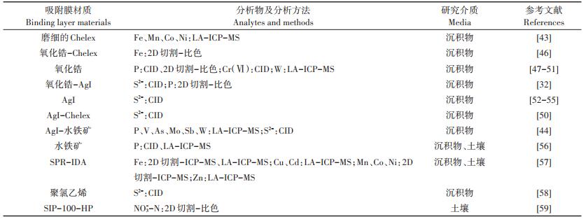
DGT已广泛用于研究SWI处元素的二维分布特征。早在1997年,Davison等[7]即利用DGT技术结合PIXE分析在100 µm的分辨率下对溪流表层沉积物和上覆微生物席(Microbial mat)及水的界面处Zn、Fe、Mn和As的二维分布进行了成像。通过对SWI处S2-和Fe的二维成像分析得到了两者重叠的“热区”(Hot spot)分布,这也是首次在海洋沉积物中研究S和痕量金属的共释放行为[55]。铁氧化物还原溶解过程中的P释放是湖泊沉积物内源P负荷的主要形成机制,DGT技术结合CID分析可以捕捉微生物活动造成硫化物微区间隙水中高浓度S2-的二维分布特征,S2-峰值通量大的硫化物微区促使了P、Fe和S的耦合反应及P的释放,原理在于间隙水中高浓度的S2-引起了FeS沉淀的生成,降低了间隙水中Fe2+浓度,促进固相铁氧化物还原性溶解进而促成P的释放[52]。与无定形Fe的氧化物或水合物结合的P是活性无机P的重要来源之一,氧化锆DGT可用于二维P分布成像[60],而氧化锆-Chelex DGT用于P和Fe同步成像,使用DGT所测得的活化态的P/Fe比值较其总量比值更加能说明铁氧化物对P释放的影响[47]。在不同季节对SWI处的活性P进行二维成像发现,夏季P的浓度高且空间分布变化大,冬季浓度低且分布相对均匀,春秋两季活性P浓度及其分布变化则介于夏冬两季之间[48]。DGT技术与LA-ICP-MS技术结合,可以高分辨同步测定多种目标物。Wu等[61]利用SPR-IDA DGT技术结合LA-ICP-MS分析,通过元素浓度的线性关系得到了Fe与S及Cu、Zn、Pb、Cd等痕量金属存在共释放现象的结论。Stockdale等[62]利用沉淀状水铁矿DGT技术结合LA-ICP-MS分析获得了SWI处P、V和As浓度的二维高分辨定量成像,而对于Mo、Sb、W和U则只得到了计数值的二维高分辨定性图像。Guan等[41]使用沉淀状氧化锆DGT结合LA-ICPMS分析获得了SWI处W浓度的二维高分辨定量成像,发现在上覆水和界面下方8 mm沉积物处均存在W的分布“热区”。
同理,DGT也用于研究沉积物介质中元素的二维分布特征。2004年Warnken等[63]首次采用SPR-IDA DGT技术结合LA-ICP-MS分析对沉积物中的Co进行二维分布成像分析,图像分辨率达100 μm,该研究为后续对Co、Ni、Cu、Zn、Cd和Pb等阳离子型金属进行二维成像分析提供了方法学支撑。Stockdale等[44]使用AgI-水铁矿DGT结合CID或LA-ICP-MS分析可对S与P、V、As的分布进行同步测定,证实了淡水沉积物中硫酸盐还原菌活性升高与其磷酸盐摄取有关。Ding等[64]利用氧化锆DGT结合2D切割-比色分析对沉积物中的P进行二维分布成像分析,发现表层与底层沉积物的活性P(DRP)浓度更高,首次证实了底栖生物水丝蚓对沉积物局部特征的扰动影响;利用氧化锆-AgI DGT结合CID分析观测到沉积物中DRP和溶解态硫化物的共释放现象[32]。
2.3 DGT分析土壤微界面元素二维剖面信息DGT已用于研究土壤介质中元素的二维分布特征,采用沉淀状水铁矿DGT结合LA-ICP-MS分析对土壤中活化态的P进行二维分布信息的化学成像,分辨率可达到亚毫米级(水平77 μm,垂直333 μm)[56]。使用SIP-100-HP DGT结合切割-比色分析对稻田土中的硝酸盐(NO3--N)进行二维分布成像,观察到NO3--N活化的热区,意味着可以利用此方法对土壤中NO3--N的生物有效性与生物地球化学过程进行深入研究[59]。
2012年,Santner等[65]对植物根轴与根尖的局部P释放进行了成像分析,并得出了外排控制根际的最低营养浓度的结论,这是首次对土壤中植物根系的P释放进行直接的观察。2013年,Kreuzeder等[66]首次采用以氢氧化锆与SPR-IDA两种材质组成混合吸附膜,利用该类型的DGT对植物根际的P、As、Co、Cu、Mn和Zn进行二维分布成像分析,通过同时观察阴离子(P和As)和阳离子(Co、Cu、Mn和Zn)型溶质,在土壤和沉积物热点(如根际)进行溶质动力学研究,并得到了高分辨的图像。2015年,Valentinuzzi等[67]研究发现,石灰能够影响可提取元素的溶解度以及植株的吸收,通过DGT对元素的生物可利用态的二维分布成像发现了羽扇豆根际的“理想[Ca]/pH区域”,即在不受Ca毒性和微量元素(如Cd)影响的情况下仍然能够调动营养物质的区域。土壤中尤其是植物根际的二维化学成像有利于了解植物利用营养成分或吸收污染物的过程,为经济作物的种植及污染物的植物吸收提供了理论依据。
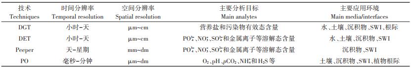
3 DGT与其他原位采样/成像技术联用研究环境微界面物质运移过程 3.1 DGT与其他原位采样/成像技术联用研究环境微界面物质运移过程的策略理论上,基于溶质扩散和吸附特性的DGT技术,可与其他一维/二维溶质浓度测定技术联用以获得多种类型溶质的同步分析,这为探究异质性环境介质中及其微界面处元素运移过程提供强大的技术支撑。DET、Peeper(包括HR-Peeper)和PO是几种常见的与DGT联用的原位采样/成像技术,这些技术的特征见表 2。商用的DET和DGT探针(板状装置)具有相同的尺寸(长×宽×厚=24 cm×4 cm×0.5 cm,底部有楔形结构),两者通常通过“背靠背”的形式插入介质中或跨过微界面,尽量保证是针对同一个剖面。当然,不可否认的是,由于环境微界面处溶质分布的高度异质性,这种方法仍无法保证被DET/DGT“劈开”的对称剖面信息完全一致。商用的HR-Peeper尺寸是固定的,自制的Peeper装置则尺寸不一,小至接近DGT探针的尺寸,大至DGT尺寸的若干倍。同理,Peeper装置和DGT探针通常也通过“背靠背”的形式插入介质中或跨过微界面。
|
|
表 2 DGT和其他原位采样/成像技术的特征比较 Table 2 Comparsions between the characteristics of DGT and other in-situ sampling/imaging techniques |
PO技术基于光致发光原理,使待测物与荧光指示剂发生作用而结合,在外部光源的激发下产生荧光信号,待测物浓度与荧光信号强度之间存在正相关性,利用特定相机捕捉荧光信号即可进行定量[11]。与DGT、DET和Peeper不同的是,PO是一种可逆的化学传感器,主要用于pH、O2、pCO2和NH4+等环境因子的成像,当前也有市场化的PO设备销售。目前为止,PO与DGT技术联用通常是在特定的培养箱(如根际箱)中进行的,方式有3种:第1种方式为在待测剖面放入PO传感膜,定期检测环境因子的分布特征;再在特定的时间点(比如植物生长的特定时期)放入DGT吸附膜,作用一段时间(通常为几小时至一天)后取出吸附膜,分析膜上富集的待测元素的分布特征;吸附膜放置期间不进行PO成像分析。第2种方式,DGT吸附膜与PO传感膜按图 3所示(以植物根际为例)组装成“三明治”式结构,放入待测剖面,其中,吸附膜更靠近剖面且与剖面之间有一层滤膜(充当DGT扩散层);吸附膜放置期间不断累积待测物质,同时进行PO成像分析;为了在DGT测定的同时获得清晰而准确的PO图像,需要尽量减少PO测定溶质(如O2)在滤膜和吸附膜中的径向扩散(Radial relaxation)效应,因此要求滤膜和吸附膜的厚度必须是超薄的(≤100 μm)[68]。第3种方式为构建DGT-PO复合膜,即将DGT吸附膜与PO传感膜“二合一”,同步测定元素和环境因子(pH、O2)的分布特征[69-70]。前两种方式发展相对成熟,而第3种方式目前仍在发展初期,需要进一步优化和完善。

|
图 3 DGT与PO技术联用研究植物根际过程示意图 Figure 3 Schematic diagram of studying plant rhizosphere process using combined DGT and PO techniques |
DGT与DET联用常用于表征沉积物生物地球化学特征,如河口沉积物中Fe(Ⅱ)与硫酸盐、磷酸盐迁移转化的相关关系、湖泊沉积物中有无蠕虫存在条件下磷酸盐热区的分布对比[13]。采用DGT与DET联用的方式对不同元素间赋存关系进行研究,在SWI处对元素一维信息采集后发现孔隙水中Fe(Ⅱ)与Sb的释放具有显著的耦合关系[71];在土壤高Cr(Ⅵ)浓度条件下对低浓度Cr(Ⅲ)进行测定,体现了DGT的预浓缩特性[72];在测定U及238U/235U同位素比值时,DGT测得的浓度较DET测得的浓度偏低[73],这种差异体现了DGT对元素生物有效态的模拟能力。
此外,DGT与DET的联用也可以用于对比环境介质中痕量金属的活化态与总量的二维分布[74],将S2-敏感膜与测量Fe2+的聚丙烯酰胺水凝胶依次贴附于玻璃板上,再附上0.2 µm的多孔PVDF(Polyvinylidene fluoride,聚偏氟乙烯)膜,制成DGT-DET结合探针,用于SWI处S2-(DGT)与Fe2+(DET)半定量化的同步测定,在界面处观察到显著的反应活性热区[58],为同一位置非单一物质的同步测定提供了依据。
3.3 DGT与Peeper技术联用研究环境微界面物质运移过程Peeper与DET都是测量溶解态元素含量的原位被动采样技术,与DET装置内的平衡相不同的是,Peeper的平衡相为溶液而非吸附凝胶。DGT与Peeper联用,可用于沉积物中生物扰动对元素释放影响的研究及元素间迁移转化的相互作用[20]。DGT与HR-Peeper联用,可以分析沉积物与其孔隙水中的活性P与溶解性P的分布与浓度变化情况[75],在对小型河流河口沉积物P、Fe、S耦合循环机制的研究中发现了涨潮落潮对该循环的重要影响[76]。
DGT与HR-Peeper常以“背靠背”的形式进行联用,分别获取有效态元素浓度的二维分布和溶解态元素浓度的一维分布,为元素活化机制的阐明提供依据。对有效态P和溶解性P进行同步测量发现随时间推移La改性膨润土对P释放的影响增强[10, 51]。应用于水稻根际时可观测到Fe2+与P的浓度衰减区域。Wang等[77]利用DGT和HR-Peeper分别测定水稻根际有效态P和溶解性P、Fe(Ⅱ)浓度的分布发现,不管是在低P还是高P土壤中,水稻吸收造成根际溶解性P和Fe(Ⅱ)的降低,DGT可以更加直观地捕捉到根际P的消耗趋势,进一步分析发现Fe在水稻根际P的稳定性调控中起主导作用。对沉积物上覆水中的溶解性Sb与活性Sb进行测定,发现Sb(Ⅲ)与Sb(Ⅴ)之间的转化决定了Sb在沉积物-水体系中的污染状况[78],而Sb的形态对湖泊中藻类的生长繁殖有极大的影响[79]。在富营养化湖泊中对Pb的溶解态与活化态进行同步测定,发现还原条件导致了Mn、Fe的溶解进而促使Pb释放至水体中,从而加剧了藻类的爆发[80],Cr在湖泊沉积物中也存在类似的季节性的迁移转化规律[81-82]。
3.4 DGT与PO技术联用研究环境微界面物质运移过程元素的有效态分布是影响其在环境中迁移转化的重要因素,而环境因子(如pH、O2)的浓度与分布以及氧化还原条件的改变与元素有效性变化息息相关,DGT与PO联用,可以得到元素与环境因子的同步成像信息。表 3中列出了DGT与PO技术联用在环境微界面研究中的应用实例。
|
|
表 3 DGT与PO技术联用在环境微界面研究中的应用(修改自Guan[6]) Table 3 Application of combined DGT and PO techniques in environmental micro-interface studies(modified from Guan[6]) |
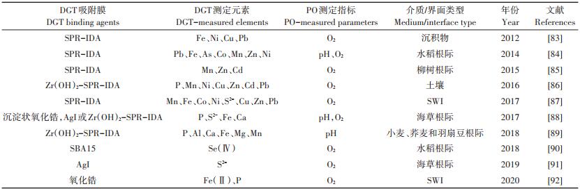
DGT与PO技术联用,研究沉积物中施加氧纳米气泡改性矿物(界面增氧材料)的锁P效果,发现该矿物通过增加SWI处O2,抑制铁矿物结合P的还原性溶出从而达到控制内源P释放的效果[93]。在SWI处将氧化锆吸附膜与荧光传感膜相结合,首次进行了SWI处活性P与O2的同步成像分析[69],通过P、Fe2+与O2的同步成像,得出了可以通过抑制P-Fe还原来抑制厌氧条件下沉积物中内源P的输入的结论[93],且Fe偶联P对活性P的潜在控制力取决于O2的供应[92],在SWI处观察到伴随植物根际氧气浓度局部急剧减少,Fe、Mn活性显著提升[87]。将DGT吸附膜与荧光传感膜结合成一张膜,能够对土壤或沉积物溶液中Mn、Ni、Co、Cd、Cu、Zn、Pb、Al和Fe离子与pH进行同步测定[70]。
在植物根际,经有机肥局部改良的土壤中P的释放与O2消耗显著增加,而P的生物有效性没有明显波动,证实了施加有机肥来增加植物根际P吸收的可行性[86]。苦草通过持续径向泌氧改变根际外围的氧化还原梯度,降低了其根际P的生物有效性[94]。海草能够通过植物自身引起的局部酸化促进其根际P的释放与Fe的还原[88],且其根际径向泌氧通过在空间、时间上限制氧化带的形成从而保护敏感的海草组织免受硫化物的入侵[91]。使用DGT-PO“三明治”装置发现在水稻根际尤其是根尖部分,伴随着O2的富集与pH的降低,As、Pb和Fe(Ⅱ)的活性显著增强,证实了水稻根际Fe的扩散与氧化的耦合对痕量金属元素的溶解、扩散及植物吸收有重要影响[84]。同样在水稻根际,Se(Ⅳ)的迁移转化能力受氧化还原条件的改变影响极大[91]。植物根际对P的活化还改变了根际土壤的pH,也不同程度地影响着Al、Fe、Mn、Ca和Mg等其他元素的活化[89]。相比对照组,根际土壤施S处理组因S的氧化所造成的根际土壤O2消耗与酸化加速了Zn、Mn和Cd的活化,表明可通过施加S修复重金属污染土壤[85]。
4 结论与展望过去二十余年的研究表明,DGT不仅在环境检测和生物有效性评价中得到了广泛的应用,还在环境微界面物质运移过程研究中显示了强大的潜力。DGT技术可以在毫米尺度研究营养盐和污染物在土壤和沉积物微界面的一维浓度分布特征。DGT技术的重要优势表现在二维化学分布成像,表征P和金属时的空间分辨率可达几十微米。近几年的一个重要发展趋势是,将DGT与DET、Peeper和PO等技术联用同步获取多种溶质的一维和二维浓度分布信息,这助推了土壤和沉积物中营养盐和污染物的生物地球化学过程的机制研究。高分辨吸附膜的不断涌现、吸附膜分析技术的多元化和分析成本的低廉化以及其他成像技术的联合使用进一步凸显了DGT在微界面过程研究中的优势,未来还需在以下几个方面继续开展工作:
(1)DGT多用于土壤和淡水系统微界面过程研究,对海洋和滨海系统微界面过程研究仍有待加强。现有部分的高分辨吸附膜可耐受较高的环境盐度,但仍有必要进一步发展耐高盐度的高分辨吸附膜和DGT原位应用辅助设施。
(2)DGT主要针对P、重金属、类金属和S2-等开展微界面过程研究,对于有机类污染物和氮素(如NO3-、NH4+)等的微界面过程研究仍然较少,需要进一步开发高选择性的吸附膜以及优化后续化学分析方法。
(3)吸附膜的均匀性是决定测量结果的准确性和分辨率的重要因素。随着DGT技术的发展,各种不同材料的特异性吸附膜应运而生。为进行环境介质中不同元素/物质的多元同步分析,含有两种及以上材料的复合吸附膜已得到开发和利用,但如何保证两种/多种吸附材料均在吸附膜中均匀分布是未来研究中需要关注的重点之一。
(4)DGT已与DET或Peeper技术联用以探究元素间的迁移转化关系,与PO技术联用以进行元素与环境因子(O2和pH)的同步分析。未来,DGT技术还可以与测定CO2、NH4+和H2S等的PO技术联用,多方位研究元素与环境因子的同步变化,进而推导微区或跨界面元素运移的驱动因子。此外,DGT还有望与其他化学成像技术(如测定二维酶活性的土壤原位酶谱技术)以及物理成像技术(如测定土壤孔隙率和根系三维分布的微型计算机断层扫描技术)联用,多角度研究环境微界面物质运移的主导因子和动力学过程。
| [1] |
曲久辉, 贺泓, 刘会娟. 典型环境微界面及其对污染物环境行为的影响[J]. 环境科学学报, 2009, 29(1): 2-10. QU Jiu-hui, HE Hong, LIU Hui-juan. Typical environmental micro-interfaces and its effects on environmental behaviors of pollutants[J]. Acta Scientiae Circumstantiae, 2009, 29(1): 2-10. DOI:10.3321/j.issn:0253-2468.2009.01.002 |
| [2] |
汤鸿霄. 环境纳米污染物与微界面水质过程[J]. 环境科学学报, 2003, 23(2): 146-155. TANG Hong-xiao. Environmental Nano-Pollutants(ENP)and their micro-interfacial processes on the aquatic particles[J]. Acta Scientiae Circumstantiae, 2003, 23(2): 146-155. DOI:10.3321/j.issn:0253-2468.2003.02.002 |
| [3] |
朱永官. 土壤-植物系统中的微界面过程及其生态环境效应[J]. 环境科学学报, 2003, 23(2): 205-210. ZHU Yong-guan. Micro-interfacial processes in soil-plant systems and their environmental impacts[J]. Acta Scientiae Circumstantiae, 2003, 23(2): 205-210. DOI:10.3321/j.issn:0253-2468.2003.02.010 |
| [4] |
Davison W, Zhang H. In-situ speciation measurements of trace components in natural-waters using thin-film gels[J]. Nature, 1994, 367(6463): 546-548. |
| [5] |
罗军, 王晓蓉, 张昊, 等. 梯度扩散薄膜技术(DGT)的理论及其在环境中的应用Ⅰ:工作原理、特性与在土壤中的应用[J]. 农业环境科学学报, 2011, 30(2): 205-213. LUO Jun, WANG Xiao-rong, ZHANG Hao, et al. Theory and application of diffusive gradients in thin films(DGT)in soils[J]. Journal of Agro-Environment Science, 2011, 30(2): 205-213. |
| [6] |
Guan D X. Diffusive gradients in thin-films(DGT):An effective and simple tool for assessing contaminant bioavailability in waters, soils and sediments[M]. Oxford: Elsevier, 2019: 111-124.
|
| [7] |
Davison W, Fones G R, Grime G W. Dissolved metals in surface sediment and a microbial mat at 100-μm resolution[J]. Nature, 1997, 387(6636): 885-888. DOI:10.1038/43147 |
| [8] |
Wang Y, Ding S, Ren M, et al. Enhanced DGT capability for measurements of multiple types of analytes using synergistic effects among different binding agents[J]. Science of the Total Environment, 2019, 657: 446-456. DOI:10.1016/j.scitotenv.2018.12.016 |
| [9] |
Davison W, Grime G W, Morgan J A W, et al. Distribution of dissolved iron in sediment pore waters at submillimetre resolution[J]. Nature, 1991, 352(6333): 323-325. DOI:10.1038/352323a0 |
| [10] |
Sun Q, Lin J, Cao J X, et al. A new method to overall immobilization of phosphorus in sediments through combined application of capping and oxidizing agents[J]. Science of the Total Environment, 2019, 694: 133770. DOI:10.1016/j.scitotenv.2019.133770 |
| [11] |
Li C, Ding S, Yang L, et al. Planar optode:A two-dimensional imaging technique for studying spatial-temporal dynamics of solutes in sediment and soil[J]. Earth-Science Reviews, 2019, 197: 102916. DOI:10.1016/j.earscirev.2019.102916 |
| [12] |
Oburger E, Schmidt H. New methods to unravel rhizosphere processes[J]. Trends in Plant Science, 2016, 21(3): 243-255. |
| [13] |
Santner J, Larsen M, Kreuzeder A, et al. Two decades of chemical imaging of solutes in sediments and soils:A review[J]. Analytica Chimica Acta, 2015, 878: 9-42. DOI:10.1016/j.aca.2015.02.006 |
| [14] |
魏天娇, 管冬兴, 方文, 等. 梯度扩散薄膜技术(DGT)的理论及其在环境中的应用Ⅲ——植物有效性评价的理论基础与应用潜力[J]. 农业环境科学学报, 2018, 37(5): 841-849. WEI Tian-jiao, GUAN Dong-xing, FANG Wen, et al. Theory and application of diffusive gradients in thin films(DGT)in the environment Ⅲ:Theoretical basis and application potential in phytoavailability assessment[J]. Journal of Agro-Environment Science, 2018, 37(5): 841-849. |
| [15] |
李财, 任明漪, 石丹, 等. 薄膜扩散梯度(DGT)——技术进展及展望[J]. 农业环境科学学报, 2018, 37(12): 2613-2628. LI Cai, REN Ming-yi, SHI Dan, et al. Diffusive gradients in thin films (DGT):Progress and prospect[J]. Journal of Agro-Environment Science, 2018, 37(12): 2613-2628. DOI:10.11654/jaes.2018-1403 |
| [16] |
Eismann C E, Menegário A A, Gemeiner H, et al. Predicting trace metal exposure in aquatic ecosystems:Evaluating DGT as a biomonitoring tool[J]. Exposure and Health, 2020, 12(1): 19-31. |
| [17] |
Davison W, Grime G W, Morgan J A W, et al. Distribution of dissolved iron in sediment pore waters at submillimetre resolution[J]. Nature, 1991, 352(6333): 323-325. DOI:10.1038/352323a0 |
| [18] |
Xu D, Chen Y, Ding S, et al. Diffusive gradients in thin films technique equipped with a mixed binding gel for simultaneous measurements of dissolved reactive phosphorus and dissolved iron[J]. Environmental Science & Technology, 2013, 47(18): 10477-10484. |
| [19] |
Gillan D C, Baeyens W, Bechara R, et al. Links between bacterial communities in marine sediments and trace metal geochemistry as measured by in situ DET/DGT approaches[J]. Marine Pollution Bulletin, 2012, 64(2): 353-362. |
| [20] |
Li C, Ding S, Yang L, et al. Diffusive gradients in thin films:Devices, materials and applications[J]. Environmental Chemistry Letters, 2019, 17(2): 801-831. |
| [21] |
Ardelan M V, Steinnes E, Lierhagen S, et al. Effects of experimental CO2 leakage on solubility and transport of seven trace metals in seawater and sediment[J]. Science of the Total Environment, 2009, 407(24): 6255-6266. DOI:10.1016/j.scitotenv.2009.09.004 |
| [22] |
Zhang H, Davison W, Mortimer R J G, et al. Localised remobilization of metals in a marine sediment[J]. Science of the Total Environment, 2002, 296(1/2/3): 175-187. |
| [23] |
Diviš P, Leermakers M, Dočekalová H, et al. Mercury depth profiles in river and marine sediments measured by the diffusive gradients in thin films technique with two different specific resins[J]. Analytical and Bioanalytical Chemistry, 2005, 382(7): 1715-1719. DOI:10.1007/s00216-005-3360-8 |
| [24] |
Clarisse O, Dimock B, Hintelmann H, et al. Predicting net mercury methylation in sediments using diffusive gradient in thin films measurements[J]. Environmental Science & Technology, 2011, 45(4): 1506-1512. |
| [25] |
Ren M, Wang Y, Ding S, et al. Development of a new diffusive gradient in the thin film(DGT)method for the simultaneous measurement of CH3Hg+ and Hg2+[J]. New Journal of Chemistry, 2018, 42(10): 7976-7983. DOI:10.1039/C8NJ00211H |
| [26] |
Zhang H, Davison W, Miller S, et al. In situ high resolution measurements of fluxes of Ni, Cu, Fe, and Mn and concentrations of Zn and Cd in porewaters by DGT[J]. Geochimca et Cosmochimca Acta, 1995, 59(20): 4181-4192. DOI:10.1016/0016-7037(95)00293-9 |
| [27] |
Wu Z, Jiao L, Wang S, et al. Multi-metals measured at sediment-water interface(SWI)by diffusive gradients in thin films(DGT)technique for geochemical research[J]. Archives of Environmental Contamination and Toxicology, 2016, 70(2): 429-437. DOI:10.1007/s00244-015-0184-1 |
| [28] |
Ding S, Wang Y, Wang D, et al. In situ, high-resolution evidence for iron-coupled mobilization of phosphorus in sediments[J]. Scientific Reports, 2016, 6: 24341. DOI:10.1038/srep24341 |
| [29] |
Han C, Ding S, Yao L, et al. Dynamics of phosphorus-iron-sulfur at the sediment-water interface influenced by algae blooms decomposition[J]. Journal of Hazardous Materials, 2015, 300: 329-337. DOI:10.1016/j.jhazmat.2015.07.009 |
| [30] |
Yin H, Kong M, Han M, et al. Influence of sediment resuspension on the efficacy of geoengineering materials in the control of internal phosphorous loading from shallow eutrophic lakes[J]. Environmental Pollution, 2016, 219: 568-579. DOI:10.1016/j.envpol.2016.06.011 |
| [31] |
Xu D, Ding S, Sun Q, et al. Evaluation of in situ capping with clean soils to control phosphate release from sediments[J]. Science of the Total Environment, 2012, 438: 334-341. DOI:10.1016/j.scitotenv.2012.08.053 |
| [32] |
Ding S, Sun Q, Xu D, et al. High-resolution simultaneous measurements of dissolved reactive phosphorus and dissolved sulfide:The first observation of their simultaneous release in sediments[J]. Environmental Science & Technology, 2012, 46(15): 8297-8304. |
| [33] |
Huang J, Bennett W W, Welsh D T, et al. "Diffusive gradients in thin films" techniques provide representative time-weighted average measurements of inorganic nutrients in dynamic freshwater systems[J]. Environmental Science & Technology, 2016, 50(24): 13446-13454. |
| [34] |
Ren M, Ding S, Shi D, et al. A new DGT technique comprised in a hybrid sensor for the simultaneous measurement of ammonium, nitrate, phosphorus and dissolved oxygen[J]. Science of the Total Environment, 2020, 725: 138447. DOI:10.1016/j.scitotenv.2020.138447 |
| [35] |
Gao Y, Baeyens W, De Galan S, et al. Mobility of radium and trace metals in sediments of the Winterbeek:Application of sequential extraction and DGT techniques[J]. Environmental Pollution, 2010, 158(7): 2439-2345. DOI:10.1016/j.envpol.2010.03.022 |
| [36] |
Luo J, Zhang H, Davison W, et al. Localised mobilisation of metals, as measured by diffusive gradients in thin-films, in soil historically treated with sewage sludge[J]. Chemosphere, 2013, 90(2): 464-470. DOI:10.1016/j.chemosphere.2012.07.064 |
| [37] |
Wang X, Huang R, Li L, et al. Arsenic removal from flooded paddy soil with spontaneous hygrophyte markedly attenuates rice grain arsenic[J]. Environment International, 2019, 133: 1051-1059. |
| [38] |
Yuan Y, Ding S, Wang Y, et al. Simultaneous measurement of fifteen rare earth elements using diffusive gradients in thin films[J]. Analytica Chimica Acta, 2018, 1031: 98-107. DOI:10.1016/j.aca.2018.05.067 |
| [39] |
Galhardi J A, Leles B P, de Mello J W V, et al. Bioavailability of trace metals and rare earth elements(REE)from the tropical soils of a coal mining area[J]. Science of the Total Environment, 2020, 717: 134484. DOI:10.1016/j.scitotenv.2019.134484 |
| [40] |
Guan D X, Williams P N, Luo J, et al. Novel precipitated zirconiabased DGT technique for high-resolution imaging of oxyanions in waters and sediments[J]. Environmental Science & Technology, 2015, 49(6): 3653-3661. |
| [41] |
Guan D X, Williams P N, Xu H C, et al. High-resolution measurement and mapping of tungstate in waters, soils and sediments using the low-disturbance DGT sampling technique[J]. Journal of Hazardous Materials, 2016, 316: 69-76. DOI:10.1016/j.jhazmat.2016.05.026 |
| [42] |
Warnken K W, Zhang H, Davison W. Performance characteristics of suspended particulate reagent-iminodiacetate as a binding agent for diffusive gradients in thin films[J]. Analytica Chimica Acta, 2004, 508(1): 41-51. DOI:10.1016/j.aca.2003.11.051 |
| [43] |
Zhou C, Van de Velde S, Baeyens W, et al. Comparison of Chelex based resins in diffusive gradients in thin-film for high resolution assessment of metals[J]. Talanta, 2018, 186: 397-405. DOI:10.1016/j.talanta.2018.04.085 |
| [44] |
Stockdale A, Davison W, Zhang H. High-resolution two-dimensional quantitative analysis of phosphorus, vanadium and arsenic, and qualitative analysis of sulfide, in a freshwater sediment[J]. Environmental Chemistry, 2008, 5(2): 143-149. |
| [45] |
Luo J, Zhang H, Santner J, et al. Performance characteristics of diffusive gradients in thin films equipped with a binding gel layer containing precipitated ferrihydrite for measuring arsenic(Ⅴ), selenium (Ⅵ), vanadium(Ⅴ), and antimony(Ⅴ)[J]. Anaytical Chemistry, 2010, 82: 8903-8909. DOI:10.1021/ac101676w |
| [46] |
孙清清, 陈敬安, 王敬富, 等. 阿哈水库沉积物-水界面磷、铁、硫高分辨率空间分布特征[J]. 环境科学, 2017, 38(7): 2810-2818. SUN Qing-qing, CHEN Jing-an, WANG Jing-fu, et al. High-resolution distribution characteristics of phosphorous, iron and sulfur across the sediment-water interface of Aha reservoir[J]. Environmental Science, 2017, 38(7): 2810-2818. |
| [47] |
Gao Y, Liang T, Tian S, et al. High-resolution imaging of labile phosphorus and its relationship with iron redox state in lake sediments[J]. Environmental Pollution, 2016, 219: 466-474. DOI:10.1016/j.envpol.2016.05.053 |
| [48] |
Meng Y, Ding S, Gong M, et al. Submillimeter-scale heterogeneity of labile phosphorus in sediments characterized by diffusive gradients in thin films and spatial analysis[J]. Chemosphere, 2018, 194: 614-621. DOI:10.1016/j.chemosphere.2017.11.178 |
| [49] |
Wang J, Chen J, Ding S, et al. Effects of temperature on phosphorus release in sediments of Hongfeng Lake, southwest China:An experimental study using diffusive gradients in thin-films(DGT)technique[J]. Environmental Earth Sciences, 2015, 74(7): 5885-5894. |
| [50] |
Wu Z, Wang S, Jiao L. Geochemical behavior of metals-sulfide-phosphorus at SWI(sediment/water interface)assessed by DGT(Diffusive gradients in thin films) probes[J]. Journal of Geochemical Exploration, 2015, 156: 145-152. DOI:10.1016/j.gexplo.2015.05.005 |
| [51] |
Wang Y, Ding S, Wang D, et al. Static layer:A key to immobilization of phosphorus in sediments amended with lanthanum modified bentonite (Phoslock®)[J]. Chemical Engineering Journal, 2017, 325: 49-58. DOI:10.1016/j.cej.2017.05.039 |
| [52] |
Wu Z, Wang S. Release mechanism and kinetic exchange for phosphorus(P)in lake sediment characterized by diffusive gradients in thin films (DGT)[J]. Journal of Hazardous Materials, 2017, 331: 36-44. DOI:10.1016/j.jhazmat.2017.02.024 |
| [53] |
Devries C R, Wang F Y. In situ two-dimensional high-resolution profiling of sulfide in sediment interstitial waters[J]. Environmental Science & Technology, 2003, 37(4): 792-797. |
| [54] |
Teasdale P R, Hayward S, Davison W. In situ, high-resolution measurement of dissolved sulfide using diffusive gradients in thin films with computer-imaging densitometry[J]. Analytical Chemistry, 1999, 71(11): 2186-2191. DOI:10.1021/ac981329u |
| [55] |
Gao Y, Van de Velde S, Williams P N, et al. Two-dimensional images of dissolved sulfide and metals in anoxic sediments by a novel diffusive gradients in thin film probe and optical scanning techniques[J]. TrAC Trends in Analytical Chemistry, 2015, 66: 63-71. DOI:10.1016/j.trac.2014.11.012 |
| [56] |
Santner J, Prohaska T, Luo J, et al. Ferrihydrite containing gel for chemical imaging of labile phosphate species in sediments and soils using diffusive gradients in thin films[J]. Analytical Chemistry, 2010, 82(18): 7668-7674. DOI:10.1021/ac101450j |
| [57] |
Tankere M S, Zhang H, Davison W, et al. Fine scale remobilisation of Fe, Mn, Co, Ni, Cu and Cd in contaminated marine sediment[J]. Marine Chemistry, 2007, 106(1/2): 192-207. |
| [58] |
Jézéquel D, Brayner R, Metzger E, et al. Two-dimensional determination of dissolved iron and sulfur species in marine sediment pore-waters by thin-film based imaging. Thau lagoon(France)[J]. Estuarine Coastal and Shelf Science, 2007, 72(3): 420-431. DOI:10.1016/j.ecss.2006.11.031 |
| [59] |
Cai C, Williams P N, Li H, et al. Development and application of the diffusive gradients in thin films technique for the measurement of nitrate in soils[J]. Analytical Chemistry, 2017, 89(2): 1178-1184. |
| [60] |
Ding S, Wang Y, Xu D, et al. Gel-based coloration technique for the submillimeter-scale imaging of labile phosphorus in sediments and soils with diffusive gradients in thin films[J]. Environmental Science & Technology, 2013, 47(14): 7821-7829. |
| [61] |
Wu Z, Wang S, Ji N. Distribution character of localized iron microniche in lake sediment microzone revealed by chemical image[J]. Environmental Science and Pollution Research, 2019, 26(35): 35704-35716. DOI:10.1007/s11356-019-06219-2 |
| [62] |
Stockdale A, Davison W, Zhang H. 2D simultaneous measurement of the oxyanions of P, V, As, Mo, Sb, W and U[J]. Journal of Environmental Monitoring, 2010, 12(4): 981-984. |
| [63] |
Warnken K W, Zhang H, Davison W. Analysis of polyacrylamide gels for trace metals using diffusive gradients in thin films and laser ablation inductively coupled plasma mass spectrometry[J]. Analytical Chemistry, 2004, 76(20): 6077-6084. DOI:10.1021/ac0400358 |
| [64] |
Ding S, Jia F, Xu D, et al. High-resolution, two-dimensional measurement of dissolved reactive phosphorus in sediments using the diffusive gradients in thin films technique in combination with a routine procedure[J]. Environmental Science & Technology, 2011, 45(22): 9680-9686. |
| [65] |
Santner J, Zhang H, Leitner D, et al. High-resolution chemical imaging of labile phosphorus in the rhizosphere of Brassica napus L. cultivars[J]. Environmental and Experimental Botany, 2012, 77: 219-226. DOI:10.1016/j.envexpbot.2011.11.026 |
| [66] |
Kreuzeder A, Santner J, Prohaska T, et al. Gel for simultaneous chemical imaging of anionic and cationic solutes using diffusive gradients in thin films[J]. Analytical Chemistry, 2013, 85(24): 12028-12036. DOI:10.1021/ac403050f |
| [67] |
Valentinuzzi F, Mimmo T, Cesco S, et al. The effect of lime on the rhizosphere processes and elemental uptake of white lupin[J]. Environmental and Experimental Botany, 2015, 118: 85-94. DOI:10.1016/j.envexpbot.2015.06.010 |
| [68] |
Lehto N J, Davison W, Zhang H. The use of ultra-thin diffusive gradients in thin-films(DGT)devices for the analysis of trace metal dynamics in soils and sediments:A measurement and modelling approach[J]. Environmental Chemistry, 2012, 9(4): 415-423. |
| [69] |
Han C, Ren J, Wang Z, et al. A novel hybrid sensor for combined imaging of dissolved oxygen and labile phosphorus flux in sediment and water[J]. Water Research, 2017, 108: 179-188. |
| [70] |
Hoefer C, Santner J, Borisov S M, et al. Integrating chemical imaging of cationic trace metal solutes and pH into a single hydrogel layer[J]. Analytica Chimica Acta, 2017, 950: 88-97. DOI:10.1016/j.aca.2016.11.004 |
| [71] |
Arsic M, Teasdale P R, Welsh D T, et al. Diffusive gradients in thin films reveals differences in antimony and arsenic mobility in a contaminated wetland sediment during an oxic-anoxic transition[J]. Environmental Science & Technology, 2018, 52(3): 1118-1127. |
| [72] |
Ernstberger H, Zhang H, Davison W. Determination of chromium speciation in natural systems using DGT[J]. Analytical and Bioanalytical Chemistry, 2002, 373(8): 873-879. |
| [73] |
Gregusova M, Docekal B. High resolution characterization of uranium in sediments by DGT and DET techniques ACA-S-12-2197[J]. Analytica Chimica Acta, 2013, 763: 50-56. DOI:10.1016/j.aca.2012.12.001 |
| [74] |
Lesven L, Gao Y, Billon G, et al. Early diagenetic processes aspects controlling the mobility of dissolved trace metals in three riverine sediment columns[J]. Science of the Total Environment, 2008, 407(1): 447-459. DOI:10.1016/j.scitotenv.2008.08.033 |
| [75] |
Yang Z, Liang T, Li K, et al. The diffusion fluxes and sediment activity of phosphorus in the sediment-water interface of Poyang Lake[J]. Journal of Freshwater Ecology, 2016, 31(4): 521-531. DOI:10.1080/02705060.2016.1181113 |
| [76] |
Pan F, Guo Z, Cai Y, et al. Cyclical patterns and(im)mobilization mechanisms of phosphorus in sediments from a small creek estuary:Evidence from in situ monthly sampling and indoor experiments[J]. Water Research, 2020, 171: 115479. |
| [77] |
Wang Y, Yuan J H, Chen H, et al. Small-scale interaction of iron and phosphorus in flooded soils with rice growth[J]. Science of the Total Environment, 2019, 669: 911-919. DOI:10.1016/j.scitotenv.2019.03.054 |
| [78] |
Ren M, Ding S, Fu Z, et al. Seasonal antimony pollution caused by high mobility of antimony in sediments:In situ evidence and mechanical interpretation[J]. Journal of Hazardous Materials, 2019, 367: 427-436. DOI:10.1016/j.jhazmat.2018.12.101 |
| [79] |
Ren M, Wang D, Ding S, et al. Seasonal mobility of antimony in sediment-water systems in algae-and macrophyte-dominated zones of Lake Taihu (China)[J]. Chemosphere, 2019, 223: 108-116. DOI:10.1016/j.chemosphere.2019.02.013 |
| [80] |
Sun Q, Lin J, Ding S, et al. A comprehensive understanding of enhanced Pb mobilization in sediments caused by algal blooms[J]. Science of the Total Environment, 2019, 691: 969-980. DOI:10.1016/j.scitotenv.2019.07.152 |
| [81] |
Fan X, Ding S, Chen M, et al. Mobility of chromium in sediments dominated by macrophytes and cyanobacteria in different zones of Lake Taihu[J]. Science of the Total Environment, 2019, 666: 994-1002. |
| [82] |
Fan X, Ding S, Chen M, et al. Peak chromium pollution in summer and winter caused by high mobility of chromium in sediment of a eutrophic lake:In situ evidence from high spatiotemporal sampling[J]. Environmental Science & Technology, 2019, 53(9): 4755-4764. |
| [83] |
Stahl H, Warnken K W, Sochaczewski L, et al. A combined sensor for simultaneous high resolution 2-D imaging of oxygen and trace metals fluxes[J]. Limnology and Oceanography:Methods, 2012, 10(5): 389-401. DOI:10.4319/lom.2012.10.389 |
| [84] |
Williams P N, Santner J, Larsen M, et al. Localized flux maxima of arsenic, lead, and iron around root apices in flooded lowland rice[J]. Environmental Science & Technology, 2014, 48(15): 8498-8506. |
| [85] |
Hoefer C, Santner J, Puschenreiter M, et al. Localized metal solubilization in the rhizosphere of salix smithiana upon sulfur application[J]. Environmental Science & Technology, 2015, 49(7): 4522-4529. |
| [86] |
Christel W, Zhu K, Hoefer C, et al. Spatiotemporal dynamics of phosphorus release, oxygen consumption and greenhouse gas emissions after localised soil amendment with organic fertilisers[J]. Science of the Total Environment, 2016, 554: 119-129. |
| [87] |
Lehto N J, Larsen M, Zhang H, et al. A mesocosm study of oxygen and trace metal dynamics in sediment microniches of reactive organic material[J]. Scientific Reports, 2017, 7: 11369. DOI:10.1038/s41598-017-10179-3 |
| [88] |
Brodersen K E, Koren K, Mosshammer M, et al. Seagrass-mediated phosphorus and iron solubilization in tropical sediments[J]. Environmental Science & Technology, 2017, 51(24): 14155-14163. |
| [89] |
Kreuzeder A, Santner J, Scharsching V, et al. In situ observation of localized, sub-mm scale changes of phosphorus biogeochemistry in the rhizosphere[J]. Plant and Soil, 2018, 424(1/2): 573-589. |
| [90] |
Shi X, Fang W, Tang N, et al. In situ selective measurement of SeⅣ in waters and soils:Diffusive gradients in thin-films with bi-functionalized silica nanoparticles[J]. Environmental Science & Technology, 2018, 52(24): 14140-14148. |
| [91] |
Martin B C, Bougoure J, Ryan M H, et al. Oxygen loss from seagrass roots coincides with colonisation of sulphide-oxidising cable bacteria and reduces sulphide stress[J]. ISME Journal, 2019, 13(3): 707-719. DOI:10.1038/s41396-018-0308-5 |
| [92] |
Yuan H, Tai Z, Li Q, et al. In-situ, high-resolution evidence from water-sediment interface for significant role of iron bound phosphorus in eutrophic lake[J]. Science of the Total Environment, 2019, 706: 136040. |
| [93] |
Yu P, Wang J, Chen J, et al. Successful control of phosphorus release from sediments using oxygen nano-bubble-modified minerals[J]. Science of the Total Environment, 2019, 663: 654-661. DOI:10.1016/j.scitotenv.2019.01.265 |
| [94] |
Han C, Ren J, Wang Z, et al. Characterization of phosphorus availability in response to radial oxygen losses in the rhizosphere of Vallisneria spiralis[J]. Chemosphere, 2018, 208: 740-748. DOI:10.1016/j.chemosphere.2018.05.180 |